Megjegyzés rétegek
A CT-megjelenítő több különböző réteget jelenít meg a betöltött képeken, amelyek hasznos információkat nyújtanak a vizsgálatról, a képről és a szeletelő orientációjáról, valamint a HU pixelértékekről és a kurzor pozíciójáról.
Vizsgálat és beteg információk
Alapértelmezés szerint a VisioVIEW a kapcsolódó vizsgálati és betegadatokat a CT-megjelenítő összes aktív képének Axiális nézetablakának bal felső és jobb felső sarkában jeleníti meg.
A megjegyzésekről és azok testreszabásáról részletes információ található a következő szakaszban a normál VisioVIEW Megjelenítőben.
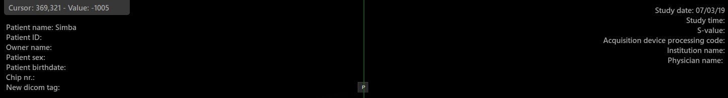
Kurzor és keresztközéppont pozíciója
CT-modalitású volumetrikus DICOM adatokkal dolgozva minden pixel HU (Hounsfield Unit) értéke a 3D testben kiemelkedő fontosságú. A Hounsfield Unit egy mennyiségi skála, amelyet az egyes szövetek radiodenzitásának mérésére használnak.
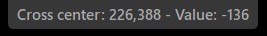
Helyezze az egérkurzort a képekre az elérhető három sík bármelyikén, hogy leolvassa az adott kurzorpozíción lévő HU értéket. Ha a kurzor nincs a kép határain belül, a két szeletelő metszéspontjának keresztközéppontja és annak HU értéke jelenik meg.

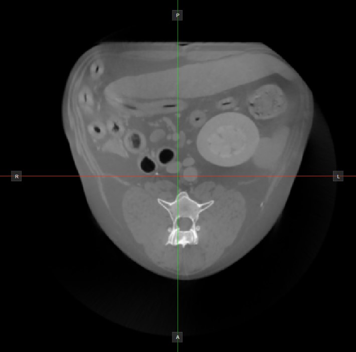
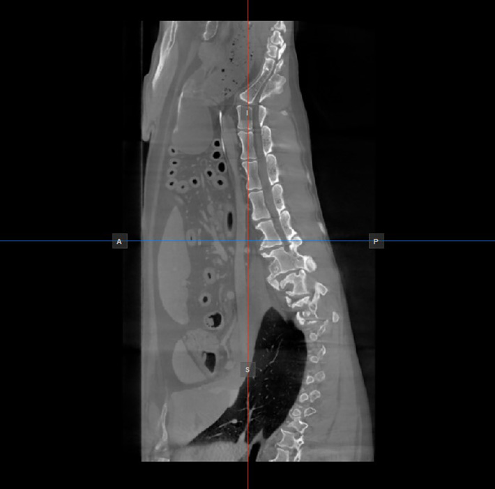
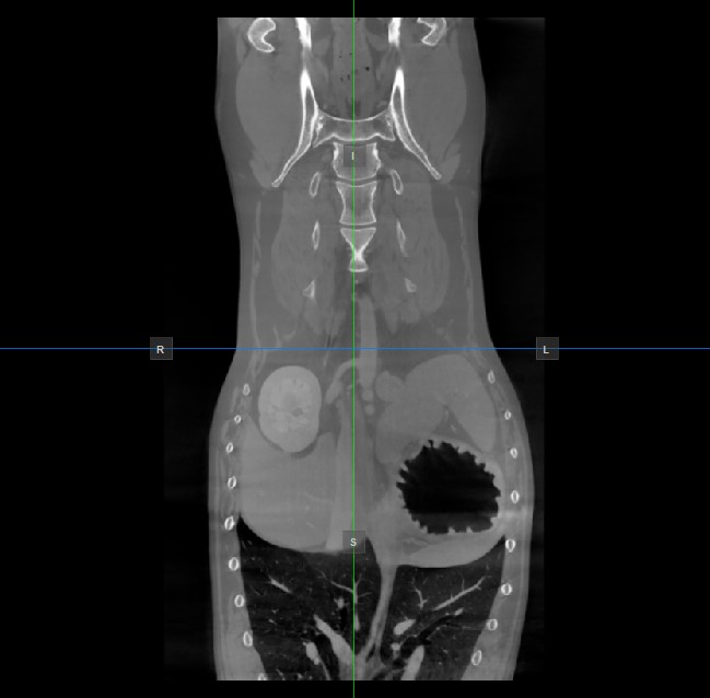
Kép orientációja és kép síkjai
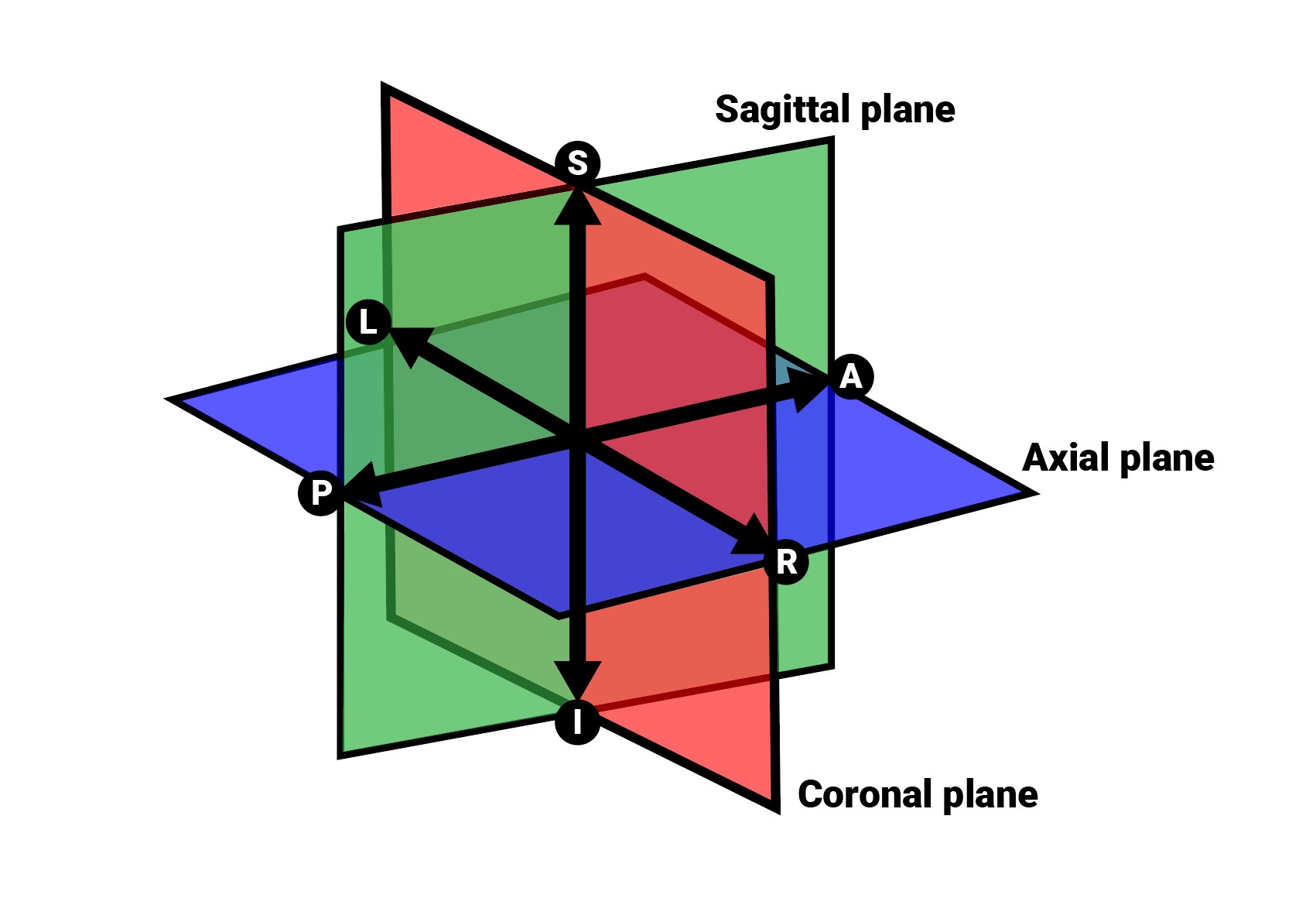
Az aktív nézetablakban nem látható másik két kép síkjának helyzetét és metszését két függőleges és vízszintes vonal jelzi, amelyeket szeletelőknek nevezünk. Minden szeletelő végpontját egy betű jelöli, amely a 3D térfogat megfelelő irányának/oldalának felel meg az RAS koordinátarendszer alapján.
Az elérhető hat oldalból csak négy van jelölve minden 2D nézetablakon. A címkék és a hozzájuk tartozó oldalak a következők:
Beteg jobb oldala (R), a test jobb oldalát jelöli
Beteg bal oldala (L), a test bal oldalát jelöli
Elülső (A), a test elülső oldalát jelöli
Hátsó (P), a test hátsó oldalát jelöli
Felső (S), a test felső oldalát jelöli
Alsó (I), a test alsó oldalát jelöli